Foot problems in elephants under human care are frequently seen. Several anatomical structures can be involved in foot problems: the sole (pad or slipper), the nails, the joint ligaments, the tendons, the distal phalanges and the joints. Many of the problems are management-related. Abnormal wear, inappropriate substrate, stereotypic repetitive movements and lack of space to move around are some of the major causes of orthopedic problems in elephant feet.
A separate chapter on regular foot care in elephants under human care can be found here.
An elephant suffering of foot problems will often demonstrate one or more of the following symptoms:
-
Swelling, pain
-
Lameness, reluctance to move
-
Black tracts on or beneath nails
-
Discharge / bad odor
-
Nail cracks
-
Overgrowth / over-wear (nails and/or sole)
-
Dry or overgrown cuticles
Examination of the foot
If the animal is well trained, it should lift its legs to allow inspection of the nails and pad of each foot. Alternatively the feet can be examined with the elephant in lateral recumbency.
Check the nail cuticles. Remember that elephants have sweat glands in their cuticles. Cuticles should not be too short as this may facilitate the invasion of microorganisms. However, they should also not be too long. Elephants seem to care for their nails by rubbing them along hard objects or their own legs. Overgrown cuticles may result in accumulation of sweat: when trimmed or when pressure is exerted on the cuticles, watery fluid may be discharged (see video).
The nails and the sole are interconnected by a structure that is an equivalent of the white area in horses and cattle. It forms a delicate connection, where microbes can penetrate into deeper layers to cause an infection (pododermatitis) ultimately resulting in osteitis and arthritis of the interphalangeal joints.
The nails should never be longer than the pad, as they should not bear weigth when the elephant is standing still nor during walking (see video). Long nails can easily develop a tear, giving access to microbes.
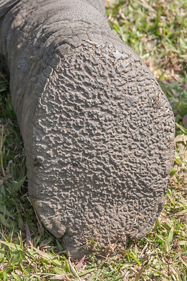
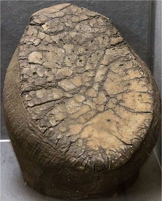
The pad of the sole must be thick and have a distinct pattern of grooves. When wear is insufficient, the grooves may become too deep and form an easy entrance for stones, dirt and microbes, which may result in a sole abscess. Too much wear results in a smooth surface and a thin pad, vulnerable for deeper sole lesions.

African elephant walking in Namibian desert (slow motion). Note how the pad and the nails form one supporting surface. The nails are not used for support. (Footage of BBC documentary)
Sole lesions

Normal sole of an Asian elephant kept in a zoo

Normal sole of an Asian elephant semi-free ranging in the forest.

Normal sole preparation of a free-ranging African elephant (Kruger National Park, South Africa)
In order to maintain a healthy sole and nails, there should be a balance between wear and (re)growth of sole and nail tissues. The main factors that determine the wear are the type of substrate, activity of the elephant and the humidity.
The EAZA Best Practice Guidelines for Elephants recommend:
Indoor and outdoor substrates should:
1. Provide choice to the elephant, allowing elephants to explore and investigate a range of substrates within their enclosure. This can provide increased activity and the cognitive benefits of decision-making.
2. Provide a degree of flexibility and accommodate the vast body mass of an elephant. Flexible ‘soft substrate’ works to absorb impact, easing the pressure on the joints and feet.
3. Provide good drainage which is beneficial for respiratory, skin and foot health. Soft substrate (such as sand) can act as a “bio floor”, allowing drainage of urine and ensuring animals are not standing in their own quite aggressive urine or lying on wet/cold floors.
4. Provide opportunities for enrichment, such as digging/hiding food which can improve musculature.
5. Allow dust bathing which provides enrichment and is beneficial for skin health.
Hard floors
Should be used only in places where long standing of elephants is not expected to occur and the elephant’s time in these areas should be minimised where possible. Concrete or rubber floors should have a non-abrasive but not smooth finish. Solid floors should be cleaned regularly and disinfected where appropriate and should provide appropriate drainage to avoid pooling of urine where elephants stand.
Sand floors
If used, type of sand should be considered. It should not be dusty, should drain well and not be able to compact (in the enclosure or gut). Sand that includes fine sand, silt or clay grades is likely to result in a dusty enclosure and compact into a solid floor. Sand grains need to be of a single size to reduce compaction. A grain with a round, rather than angular, shape will reduce compaction and will not be over wearing on the feet.
Sand depth should be of 0,8 m minimum depth, but 1,5 m is recommended. Daily maintenance of sand includes daily watering to prevent excessive dust. Sand must be regularly turned to prevent compaction and build-up of bacteria that can grow in anoxic conditions. Sand floors need to be easily accessed by specially designed heavy machinery (such as truck loaders), and purposely built concrete ramps are a must.
Where sand is retro fitted into enclosures, drains should be protected with permeable membranes and sand depths maximised where possible. Door runners should be subject to increased maintenance procedures to mitigate sand ingress. Alteration of door runners can be considered in preference to removal of substrates.
Benefits of sand include the ability to build up pillows or mounds which may be used by older animals to sleep against or aid older/arthritic elephants when getting up. Animals also can benefit from rolling/ playing in or on mounds and they can act as visual barrier. The drainage provided is beneficial to aid calves in standing up quickly post birth. Sand can be used in outdoor areas, in all weather, and can withstand extreme cold or wet weather (with appropriate drainage).
Grass
If possible (i.e. space available will not be significantly compromised with paddock rotation technique), allow grazing access for elephants, which prolongs foraging times.
Under improper environmental conditions the following sole lesions can develop:
-
Figure 1. Excessive wear: too much wear results in a smooth surface and a thin pad, vulnerable for deeper sole lesions.The sole of an adult elephant should be at least 20 mm thick.
-
Figure 2. Insufficient wear: the grooves may become too deep and form pockets, which are an easy entrance site for stones, dirt and microbes and can lead to a sole abscess.
-
Figure 3. Crack/tear
-
Figure 4. Sole fistula/ulcer/abscess
-
Figure 5. Sole detachment (partial or complete)
-
Result from primary infection (cowpox and Foot-and-mouth disease)
-
Secondary infection after trauma, moisture, weight overload, dirt:
-

Figure 1. The sole of this zoo-kept African elephant is too thin and health a smooth surface, making it vulnerable to cracks, perforations and nail lesions. Note the small cracks in the nails.

Figure 2. Overgrown pad and nails. See also: International Elephant Foundation.

Figure 3. Crack in sole of African elephant kept on a concrete floor

Figure 4. Fistula/ulcer/abscess in the sole of an Asian elephant spending most of the time on the streets in Laos.

Figure 5. Partial sole detachment in an Asian elephant orphan at the Dak Lak Elephant Conservation Center (Vietnam), caused by keeping him on a wet floor (rainy season).


Injuries to the sole of the foot are particularly difficult to manage because it is hard to keep them clean and prevent infection. Careful pedicure of the sole may reveal bruces in the soft horn tissue, that resulted from abnormal local pressure on the sole. (Courtesy: Susan Mikota).

These bleedings are blood lines in the horn lamellae (not to be confused with sole bruses) that may be seen when abnormal forces have been excerted on the nail (e.g. too long nail) (Courtesy: Susan Mikota).
Perforated, infected sole lesions due to trauma (Courtesy: Susan Mikota)

Partial pad and nail loss in a 54-yrs-old female Asian elephant. Click here to read the case report.
Foreign body pad perforation
One case report describes the treatment of a pad abscess resulting from the penetration of a wire through the pad in a 19-yr-old female Asian elephant (Elephas maximus) housed at the Paris Zoo (Ollivet-Courtois 2003). The cow presented with acute right forelimb lameness and swelling that persisted despite 4 days of anti-inflammatory therapy. Under anesthesia, a 10 x 0.5 x 0.5 cm wire was extracted from the sole of the right foot. There was a 2-cm-deep, 7-cm-diameter abscess pocket that was subsequently debrided. Regional digital i.v. perfusion was performed and repeated 15 days later, using cefoxitin and gentamicin on both occasions. Between treatments, the cow received trimethoprim–sulfamethoxazole and phenylbutazone orally. Within 2 days of administering anesthesia and the first perfusion treatment, the lameness improved dramatically. When phenylbutazone was discontinued 1 wk after the first treatment, the lameness had completely resolved. At the second treatment, there was no evidence of further soft tissue infection, and the abscess pocket had resolved.

A 10 x 0.5 x 0.5 cm piece of curved wire was found penetrating the right front foot of a female Asian elephant.

A rope tourniquet was placed above the
right carpus, and venous access was obtained using a 21-gauge, 0.8 mm butterfly catheter in a palmar superficial vein of the right foot to perform regional interdigital perfusion.
Complete sole detachment can be caused by:
-
Foot-and-Mouth disease (FMD)
-
Generalized Cowpox infection.
Foot and Mouth disease
Both elephant species are susceptible to FMD. However, severe disease has been reported (anecdotically) more in Asian elephants than in African elephants. A description of FMD in African elephants after experimental (!) infection is found here (Howell, 1973). In range countries it is important to avoid direct and indirect contact between cattle and elephants, especially during FMD-outbreaks in the cattle population. Vaccination of cattle is important to reduce the risk of FMD to elephants living in the same area as cattle. For vaccination data, see also: The use of Vaccination of FMD in zoo animals (Schaftenaar, 2002)


Complete sole detachment in an African elephant after experimental infection with FMD-virus.
Cowpox
Both elephant species are susceptible to cowpox infections, though more severe clinical impact is seen in Asian elephants. Symptoms can range between external pox lesions on the skin to complete sole detachment, as well as internal pox lesions in various organs. Vaccination is practiced in European zoos (for vaccine information, click here).
NB: Cowpox disease is a zoonotic disease!


To reduce the pressure on a thin or perforated sole, a rubber sole can be glued on the thin sole. This material can last for 2-6 weeks when properly glued to the sole.
Avenir Light is a clean and stylish font favored by designers. It's easy on the eyes and a great go-to font for titles, paragraphs & more.
A more frequently used tool to reduce the pressure on the sole, is a sandal or boot made locally, fitting the foot of the elephant. This shoe should be removed daily to inspect and treat the injury.


Custom-made sandal, Fowler & Mikota 2006


Another example of custom-made sandals to protect an injured sole (Myanmar. Courtesy Susan Mikota)


These sandals were made by commercial companies Teva and Nike (Courtesy Susan Mikota)
Edges of the sole bordering the sole defect were thinned (Dak Lak Elephant Conservation Center, Vietnam)
Treatment of sole lesions
Excessive wear: the thin and smooth horn layer has to regrow. This means that wear has to be reduced. This can be achieved by reducing the time spent by the animal on a hard floor, changing the floor surface and substituting it by softer materials; i.e. concrete floors can be covered by an epoxy layer or deep sand. A concrete sleeping area should be replaced by a sand floor (see EAZA Best Practice Guidelines for Elephants).
The thickness of the pad can be measured by ultrasound examination.
Overgrown sole: horn can be trimmed away using a hoof knife. Usually the nails will also need trimming. The use of a drawknife is not recommended, as it easily removes too much from the sole. Make sure that the nails are always shorter than the sole!
Crack/tear: as these are usually the result of a too thin sole, measures to increase the sole thickness should always be taken. If the crack hasn't perforated the sole completely, the edges of the crack can be cut away using a hoof knife with the aim to prevent accumulation of dirt and to reduce the pressure on the thinnest part of the crack. A sandal should be considered is there is a risk of sole perforation.
Sole fistula/ulcer/abscess: if the sole is perforated, microbes will have entered the underlying tissue. Frequent pedicure will be required in order to drain the affected tissues. The edges of the fistula need to be made as thin as possible with a smooth transition to the thicker part of the sole. This will reduce the pressure on the infected area. The fistula/ulcer/abscess must be flushed daily with saline solution and a mild disinfectant. Soaking the foot in a foot bath should be considered (see images below). Several solutions have been used in elephants (see table below). Epsom salt is probably superior to the other solutions, while copper sulfate might be too caustic for this type of injury.
As long as the effluent is sufficiently drained with the help of frequent trimming, the use of systematic antibiotics is not indicated.

List of foot bath solutions as described in Fowler & Mikota 2006


Simple foot soaking bath by a well trained Asian elephant, Dak Lak Elephant Conservation Center (Vietnam)
Custom-made foot soaking bath, Laos (courtesy Dionne Slagter)
Partial sole detachment: this condition can be the result of long exposure of the sole to water and dirt. Standing on a hard floor will predispose for this lesion.
Parts of the sole that are detached from the underlying tissue must be entirely trimmed away using a hoof knife. Leaving a sole flap - even a small one - will result in extension of the infected area. The edges of the sole bordering the defect, must be made thin and smooth in order to avoid pressure of the remaining sole on the fragile exposed tissues.
Regular trimming of the edges of the sole around the defect and daily flushing and foot bath are required. In some cases the use of a sandal or boot may be needed.

Complete sole detachement: this condition is usually the result of trauma or a viral infection: Foot and Mouth Disease and Cowpox have been associated with complete sole detachment, always accompanied by other severe symptoms caused by these viruses. As there are no specific treatment options for these viral diseases, only symptomatic treatment can be given. Bandaging the affected legs has been practiced, but one should not be optimistic about the results. In some cases, humane euthanasia will be the only option to prevent the animal from suffering.
Traumatic sole detachment can be expected if the elephant has been trapped in a snare. Cleaning, disinfection (mild solution soaking foot bath) and sometimes bandaging of the affected foot will be necessary until the wound has closed. In some cases, the regenerated tissue that covers the wound is strong enough to withstand the pressure of the body weight. If that is not the case, a prosthesis will be needed to provide sufficient protection.
-
Cowpox infection in elephants. 1996. Proceedings of the annual conference of the European Association of Zoo and Wildlife Veterinarians.
-
Fowler ME 2006. Foot disorders. In: Biology, Medicine, and Surgery of Elephants. Fowler & Mikota, 271-290.
-
Howell P.G., Young E, Hedger R.S. 1973. Foot-and-mouth disease in the African elephant (Loxodonta africana). Onderstepoort J Vet Res. 1973 Jun;40(2):41-52.
-
Johnson G., Smith J., Peddie J., Peddie L., DeMarco J., Wiedner E. 2018. Use of glue-on shoes to improve conformational abnormalities in two Asian elephants (Elephas maximus). J. Zoo&Wildl Med. 49(1): 183–188, 2018.
-
Ollivet-Courtois, F., Lécu, A., Yates R.A., Spelman L.H. 2003. Treatment of a sole abscess in an Asian elephant (Elephas maximus) using regional digital intravenous perfusion. Journal of Zoo and Wildlife Medicine 34(3): 292–295, 2003
-
Schaftenaar W. 2002. Use of vaccination against foot and mouth disease in zoo animals, endangered species and exceptionally valuable animals. Rev. sci. tech. Off. int. Epiz., 2002, 21 (3), 613-623.

