Orthopedic problems
Elephants do not often show signs of lameness. Nevertheless, orthopedic problems are quite common. A survey about the causes of death in the European studbooks of African and Asian elephants over 5 years of age, revealed that in 12% and 30% respectively of the cases, orthopedic problems played a major role in the cause of death (Hess 2022).
The most frequently reported problems are related to the feet, joints and muscles. A special issue is the occurrence of metabolic bone disease in bottle-raised young elephants.
Normal features of the locomotion system


Anatomical features of the skeleton
The elephant has some special features that distinguishes them from other mammals. The long bones are massive, lacking the typical bone marrow cavities. Instead, the long bones of elephants are completely filled with dense cancellous bone, where hemopoiesis is taking place. In the standing elephant, the angles of the joints are almost straight. The neck is relatively short. Figure 1: Asian elephant (Green Hill Valley, Myanmar). Figure 2: African elephant skeleton (Veterinary Faculty Utrecht University, the Netherlands)
Foot anatomy terms
-
Front foot = fore foot = manus
-
Hind foot = rear foot = pes
-
Phalanges = toes = digits
-
Pad = sole = slipper
-
Palmar = front pad
-
Plantar = back pad
-
Carpus = wrist
-
Tarsus = ankle
-
Nail = horn wall + nail pad horn

Fat cushions
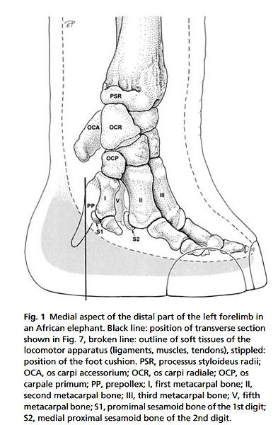
Each foot of the elephant is equipped with a large subcutaneous cushions which play an important role in distributing forces during weight bearing and in storing or absorbing mechanical forces. One study about these cushions in the African elephant was published by Weissengruber in 2006. In both the forelimb and the hindlimb a 6th ray, the prepollex or prehallux, is present. These cartilaginous rods support the metacarpal or metatarsal compartment of the cushions. None of the rays touches the ground directly. The cushions consist of sheets or strands of fibrous connective tissue forming larger metacarpal/metatarsal and digital compartments and smaller chambers which are filled with adipose tissue. The compartments are situated between tarsal, metatarsal, metacarpal bones, proximal phalanges or other structures of the locomotor apparatus covering the bones palmarly/plantarly and the thick sole skin. Within the cushions, collagen, reticulin and elastic fibres are found. In the main parts, vascular supply is good and numerous nerves course within the entire cushion. The high concentration of sensory receptors such as Vater–Pacinian corpuscles within the cushion and Meissner corpuscles in dermal papillae of the adjacent skin might rank an elephant’s foot among the most sensitive parts of its body. Together, the mechanical and sensory functions of the feet enhance the ability of elephants effectively to move through and analyse their physical environment.
The micromorphology of elephant feet cushions resembles that of digital cushions in cattle or of the foot pads in humans but not that of digital cushions in horses.

Copied illustration of the foot anatomy from Weissengruber et al., 2006 (doi: 10.1111/j.1469-7580.2006.00648.x
Normal locomotion
Elephants predominantly support on their pads (foot soles). The nails are not used to force locomotion. This is nicely demonstrated in the slow-motion video below (BBC).

During walking the head of the elephant shows minimal movements. If there is any form of lameness, especially in one of the front legs, the animal might use its head to facilitate the movement of the front leg in cranial direction. In the absence of orthopedic problems, the hind feet are placed cranial to the foot step of the front foot on the same side. This is clearly demonstrated in the slow-motion video of African elephants in the Namibian desert below (BBC) and the normal-speed video of an adult Asian elephant bull in Vietnam.


Elephants can't trot, canter, gallop or jump. They always walk in normal gait. When they walk slowly, their speed is approximately 4 km/h (2.5 miles/h). However, they can reach a speed of 25-60 km/h (16-25 miles/h) over a short distance. The hind foot is placed in the foor print of the front foot or even slight more cranial.
Normal anatomical features of the elephant foot
Usually the forefeet of the Asian elephant have 5 nails and the hind feet only 4. The African elephant has 4 nails on the forefoot and 3 on the rear one.
The weight of the body is evenly distributed over the toes by means of a thick cushion, placed between the sole and the phalanges (photo African elephant foot Kruger National Park, South Africa). The digits form a ±45° angle with the sole, as shown in the radiograph below (Fowler and Mikota 2006).


This photo shows the longitudinal section of the elephant foot with the sole, nail, phalangeal bones, cushion and tendons. Note the short distance between the nail and the distal phalangeal bone (Fowler and Mikota 2006)

The nails are numbered medial to lateral. If there are 4 nails in front they are numbered 2,3,4,5. The bones don’t change – there are always 5 digits so digit 1 is still there but there in no associated nail. In Asian elephants there are typically 4 nails on the rear foot so they are numbered 2,3,4,5.
The African elephant's toes are numbered 5,4,3,2 (front) and 5,4,3 (rear) respectively.

This diagram shows the bones of the front foot and the respective phalanges of an Asian elephant (Fowler&Mikota 2006)

This diagram shows the bones of the hind foot and the respective phalanges of an Asian elephant (Fowler&Mikota 2006)

Radiograph of the left front foot of an Asian elephant, showing the phalangeal bones P1,P2 and P3 (Fowler&Mikota 2006)
Sixth toe
The elephant has unique cartilaginous structures in the feet that are thought to have a stabilizing function. In the front foot the structure is is called a prepollex. It attaches between the first carpal bone and the first metacarpal bone and extends to the sole. In the hind foot it is called a prehallux. A recent study has claimed that this structure should be considered a sixth toe because over time the tissue becomes hard like bone.

The healthy sole

The sole (pad or slipper) of the elephant's foot is a thick cornified but flexible integumentory structure, with a surface relief that looks almost similar to the skin. It is important to respect this surface when performing pedicure. The thick sole must protect the elephant from penetrating trauma by foreign bodies. A healthy sole is maintained by providing a dry environment. Long periods in muddy and humid circumstances can lead to sole injuries and even sole detachment.
The photos show the nicely structured sole of a (dead) wild African elephant (Kruger National Park, South-Africa) and the sole of a captive Asian elephant.

The sole of the elephant foot should have a minimum thickness of 2 cm. This can be measured by ultrasound examination. Its surface should be rough with a distinct relief.
The growth of the sole epithelium is from 0.5 to 1.0 cm per month. If the sole does not wear sufficiently, it becomes thickened, and because the thickening is seldom uniform, defects are produced that lead to pocket formation and overgrowth, which sets the stage for infection.
The healthy nail
The nail consists of two parts: the wall and the sole part, which are connected at the sole side. This junction is an important area where infections can emerge if its integrity has been severed by excessive abrasion on hard floors (concrete stable, tar roads) or wrong pedicure. This connection site is comparable with the so-called 'white zone' in hoofed mammals. The white line (or white zone) structure is illustrated in the figures and photos below (Benz, 2005).

The nails should be shorter than the pad, without cracks and U-shaped. The skin in between 2 nails should be clean and flexible. When there is hyperkeratosis in this area, this may cause discomfort to the elephant as the hard hyperkeratotic tissue acts as a foreign body by pinching the interdigital skin an dirt can accumulate into the interdigital space. There should be room for at least one finger between 2 nails.
The thermographic images of a healthy nails shows a regular distribution of the temperature dispersed over the entire nail.


Like in hoofed mammals, the nails are connected with the underlying phalanges by lamellae or horn leaflets. Benz (2005) describes the different parts of the nail:
a: corial part of the horn wall: cuticle area
b: lamellae (horn leaflets)
c: white zone
d: sole horn
Cuticle and sweat glands
The cuticle of the nail is the keratinized skin at the junction with the nail. They should be soft and flexible. This is a vulnerable area as microorganisms may pass this natural barrier after (micro)trauma. The elephant seems to maintain the cuticles by rubbing them gently against objects. Elephants that are kept in moist, muddy conditions, are likely to develop problems with the cuticles. They may overgrow and become hardened when they dry, resulting in cracks and infection. During pedicure, one should be well aware of the protecting function of the cuticles and never remove more than necessary.

References
-
Benz, A. 2005. The elephant’s hoof: Macroscopic and microscopic morphology of defined locations under consideration of pathological changes. Master's thesis, Veterinary Faculty of the University Zürich, Switzerland.
-
Fowler M.E. and Mikota S.K. 2006. Biology, Medicine, and Surgery of Elephants. 271-290.
-
Hess A. 2022. Lesions found in the post-mortem reports of the Asian (Elephas maximus) and African (Loxodonta africana) elephants of the European Association of Zoos and Aquaria Master's thesis, Department of Exotic Animal and Wildlife Medicine University of Veterinary Medicine Budapest, Hungary.
-
Schiffmann C. 2021. Posture Abnormalities as Indicators of Musculoskeletal Disorders in 12 Zoo Elephants – a Visual Guide. Gajah 53 (2021) 20-29.
-
Weissengruber, G.E., Egger, G.F., Hutchinson, J.R., Groenewald, H.B., Elsässer, L., Famini, D. and Forstenpointner, G. (2006), The structure of the cushions in the feet of African elephants (Loxodonta africana). Journal of Anatomy, 209: 781-792. https://doi.org/10.1111/j.1469-7580.2006.00648.x